Switch language or website
Switch Language
Other countries and websites
AtlasPROfilax® is available around the world in many languages. Please select your region to switch to another website.
AtlasPROfilax® is available around the world in many languages. Please select your region to switch to another website.
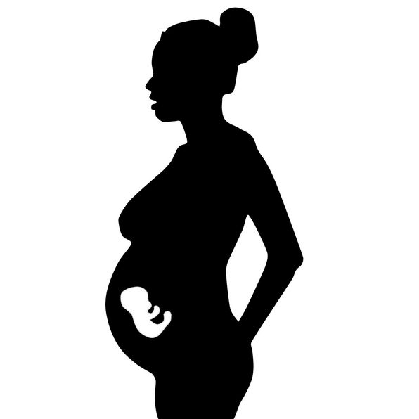
It would be unethical to double-blind the method with pregnant women because although theoretically, there is no probability of harm - as we humans are autopoetic - the reaction that may occur in the fetus is not easily calculable. For this reason, AtlasPROfilax® Academy of Switzerland has not considered it appropriate to advance research in this area.
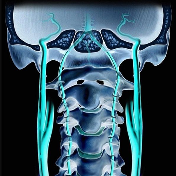
In cases of significant levels of compression on the vertebral artery on its way to the skull, an ischemia of the vertebro-basilar system can be generated, which may become evident during ipsilateral rotation and head extension movements. As a precaution, the method is not applied in cases where significant vestibular alterations are produced when the head is mobilized.
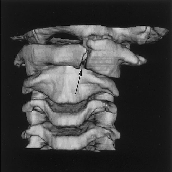
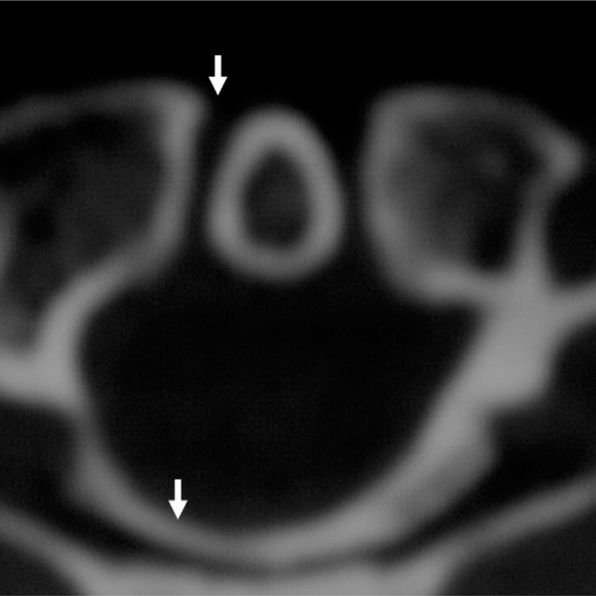
The atlas can show a fracture of the posterior arch, the anterior arch, transglenoid or both arches (anterior and posterior). Such injuries are the result of polytrauma or cranio-encephalic trauma. The approach to these cases should be strictly clinical and in some cases, surgical. In relation to the axis, the fracture by avulsion of the dens, of the base of the dens or of the axial body, should be resolved orthopedically or surgically. Under no circumstances can the suboccipital muscles be worked until the person has been stabilized and has had a complete recovery from their injuries.
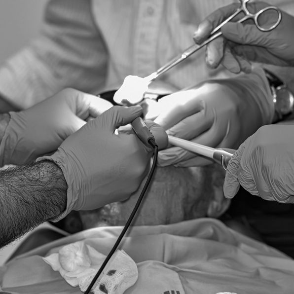
In general, any post-operative requires immediate and subsequent care that must take into account the type of intervention, type of anesthesia received, medical treatments prescribed, assessments by doctors and nurses. To this end, the best policy is caution in order to allow the body to complete the process of adjustment, adaptation and recovery. In fact, depending on the type of surgery performed, the prudential time frame for working the suboccipital muscles will vary. In some cases, months will pass and in others, surely years.
Among them, the most common are atlas occipitalization, os odontoideum, spina bifida at C1 and C2, atlanto-axial fusion and hypoplastic atlas. This type of anomaly brings with it a great risk of developing neurological alterations and is usually seen correctly through conventional radiography in a neutral position. The method will not be applied if the atlanto-axial distance is greater than 4 mm or the channel width less than 14 mm. All this is done to avoid aggravations that could compromise the physical integrity of the person.