Introduction to the AtlasPROfilax® method

AltasPROfilax® method for professionals

Technical information for health professionals about the AtlasPROfilax® method

Definition

Swiss myofascial method by tissue resonance vibropressure. It consists of a highly specific vibratory-resonant application on the short neck muscles that positively affects the cranio-cervical hinge (Occipital - Atlas - Axis) because it corrects the Minor Intervertebral Derangement of the Atlas that consists of the Condylar Compression of the Occipital and the Cranio-Caudal Deviation of the Atlas.

Hinge: Transition vertebrae between each vertebral region, characterized by the instability that gives them their greater mobility (Rothman and Simeone, 1989; Hamill and Jnutzen, 1995).

Condylar Compression Syndrome

The Occipital Condylar Compression Syndrome refers to the pressure of the occipital condyles between the upper concave articular facets of the Atlas (glenoid cavity) by the limitation caused by the basilar extradural connective tissue. This can lead to dysfunctions of the occipital, sphenobasilar and temporal scales of the skull. 

This condition, apparently harmless, can trigger speech problems, hyperactivity, vertebro-basilar headache, essential hypertension, orthostatic dizziness, irritable bowel and tension of the atlanto-occipital joint, among others. As well as some malformations at the occipital level, foramen magnum and some ducts where cranial nerves run.

The cranio-facial axis is the vertical axis that runs from the head to the feet. The cranio-flower deviation of the Atlas consists of a small parasagittal alteration of the position of the Atlas, a vertebra that in many cases is found in a descending direction with respect to the body, as a consequence of diverse mechanical factors.

Both conditions together, the Occipital Condylar Compression and the Cranio-Caudal Atlas Deviation are responsible for the irritation of the brain stem, the compression and angulation on one of the vertebral arteries, the congestion of the intracranial CSF, the poor vascularization of the superficial petrous nerve, the reactive muscular-fascial syndrome and the transfer of the center of gravity of the spine, from L2-L3 to L5-S1.

    Most frequent events

    This usually manifests itself in various ways. The most frequent are:

    • Headaches and dizziness.
    • Musculoskeletal pain disorders.
    • Coordination and balance disorders.
    • Alteration of muscle tone with the appearance of contractures.
    • Increased probability of developing lumbar disc herniations.
    • Displacement of the center of gravity of the skull and therefore of the spine.
    • Interferences towards the neurological centres that control the autonomous body functions.
    • Cervical misalignment that can be transferred to other vertebral segments, leading to scoliosis.
    • Affection of serotonin levels and with it, very possibly, decreased energy and insomnia.
    • Disorders in digestion, absorption of nutrients and elimination due to alterations in the vagus nerve pathways.
    • Hyperactivity of the sympathetic nervous system that generates immune stress: e.g. susceptibility to fungi and allergies.

    The spine

    More than a simple longitudinal axis of the body, the spine is, together with muscles and ligaments, a stabilization mechanism. It is also a point of muscle insertion and a protection system for the spinal cord. Thanks to the special gearing of vertebrae and discs, it is capable of performing movements at different amplitudes, depending on the segment to be used.

    Biomechanically, this complex osteofibrocartilaginous structure is made up of a system of three pillars. An anterior abutment for resistance and cushioning (vertebrae - intervertebral discs) and two posterior abutments for movement (interapophyseal joints and ligaments). Functionally, the plates and ligaments facilitate the function of the posterior pillars.

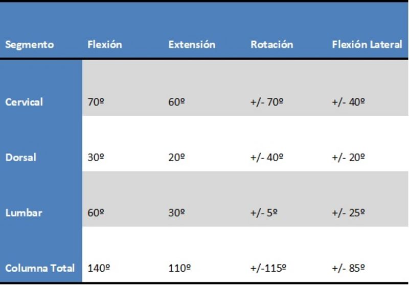
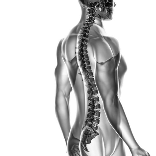
    The cervical spine has to dissipate the forces acting on the head, protect the brain stem and the nerves and blood vessels that travel through it. Its joint system has three joint systems: Anterior (intersomatic-uncovertebral) and two posterior (zygapophyseal). Its range of motion is very high and the disc diameter is relatively small.

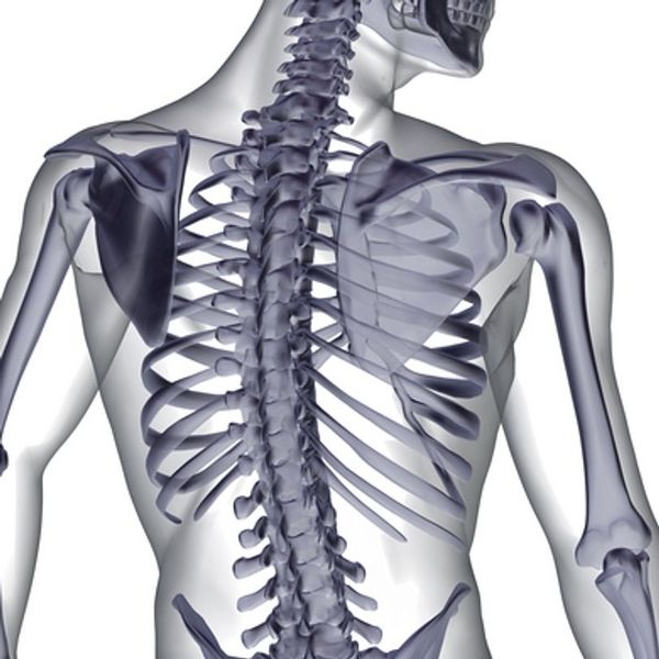
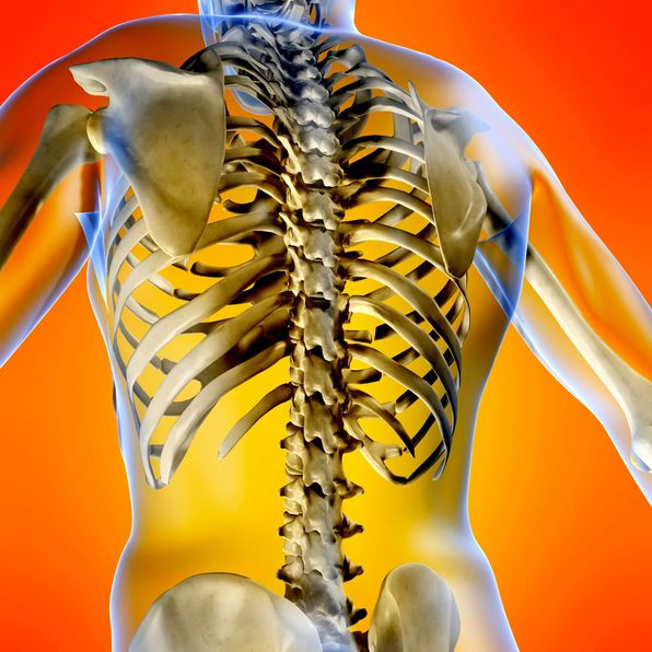
    The thoracic spine must protect the medullar axis and provide the statics of the trunk. The discs have similar height and smaller diameter than in the cervical or lumbar region. The stability of this area is given by the attachment of some dorsal vertebrae to the ribs and muscles. It is especially the yellow ligaments that resist the tensile forces of this segment.

    The lumbar spine must support the weight of the upper spine and the trunk, which in turn must transmit compressive and shear forces to the lower spine. The deformability of its discs allows for mobility. It is the joint veneers and discs that generate stability. L3 is the vertex of lumbar lordosis, L2 is the center of gravity of the spine and L5 is the transition zone to the sacrum. 

    The sacral region must influence the distribution of the lines of force of the descending myofascial chains as well as the position and mobility of all the vertebrae. In communion with the ilia, it forms the pelvic girdle which stabilizes the pelvis and transmits the load forces to the pelvic ends. It is the sacroiliac ligaments that are responsible for resisting rotation, shearing and tension in the pelvis.

    The coccyx must prolong the sacral curve, support the rectal ampulla and provide insertion for the anococcygeal ligament and the gluteus maximus and coccygeal muscles.  Its movements are flexion and extension. Lumbosacral spine disorders decrease sagittal pelvic rotation and therefore hinder coccyx flexion at seating. Its stability is based on the anterior and posterior sacrococcygeal ligaments.

    The spine and its nerve relationship to the body

    The organization of the SNP is due to the metameric division of the embryo and its transformations during the period of development. In the human being, the somites formed by mesoderm, are distributed on both sides of the neural tube to become dermatomes (skin), myotomes (skeletal muscle) and sclerotomes (vertebrae and skull). 

    Once the sclerotomes are differentiated, the caudal half must fuse with the adjacent cranial half to form vertebrae. Once the neural tube grows, the shadows and spinal nerves follow. From the neural tube the spinal cord emerges and from its outgrowth, the spinal nerves originate. Each nerve associated with the myotome and dermatome of the respective segment.

    The spinal segment is a metamere that gives sensory-motor innervation to an embryonic division. The metameres are responsible for the segmental distribution of the sensory fibers in the body. Their distribution is done in spinal segments for clinical reasons, although the spinal cord does not suffer segmentation in the embryo, only the paraxial mesoderm along the notochord does.

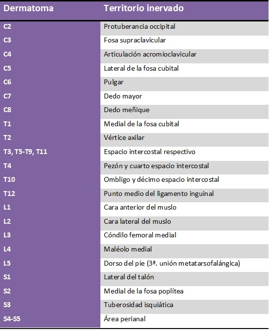
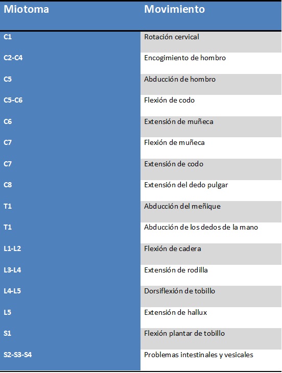
    The innervated skin area that carries sensitive information to a root is called a dermatome, the muscle group innervated by a single spinal segment is known as a myotome, and the skeletal area innervated by a segment of the spine is called a sclerotome. For each metalmere there is a segment of the spine, a dermatome, a myotome, and a sclerotome.

    As a rule, the number of dermatomes is equal to the number of spinal segments. However, C1 is the exception in that it has no cutaneous territory. Thanks to the maps that have been developed for the location of dermatomes, it is possible to make neurological diagnoses to detect spinal cord and CNS lesions.

    The Atlas Vertebra

    Because it is an incomplete vertebra, the Atlas has fused its vertebral body to give rise to the Axis dens. Its posterior tubercle represents the spinous process. The glenoid cavities and the flat veneers of the lower face of the lateral masses represent the articular processes. The posterior arch is stated as a lamina. The sides of the arch are the pedicles.

    Its two lateral masses are joined by the posterior and anterior arches. The lower face articulates with the Axis. The transverse processes originate from the outer face of the Atlas, while the inner face serves as the insertion point for the transverse ligament. The articular veneer articulates with the dens. The eminences of the Atlas are its tubercles, anterior and posterior.

    The vertebral foramen of the Atlas is anatomically limited by the lateral masses and the anterior and posterior arches. On the one hand, it contains the Axis dens and on the other hand, the bulb-medullary junction with its respective meninges. The transverse processes contain the transverse foramen through which the vertebral arteries run while the upper face of the posterior arch contains the vertebral artery and the C1 nerve.

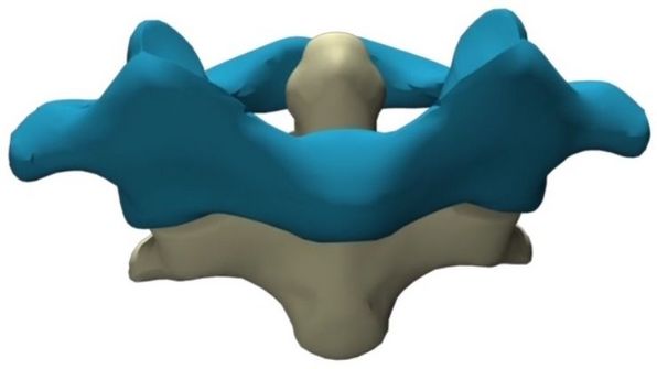
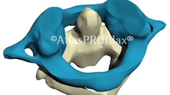
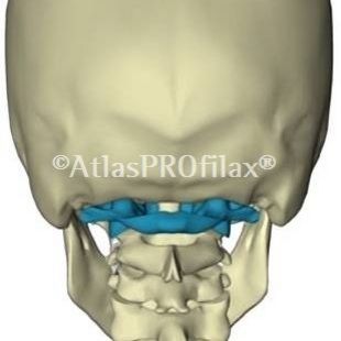
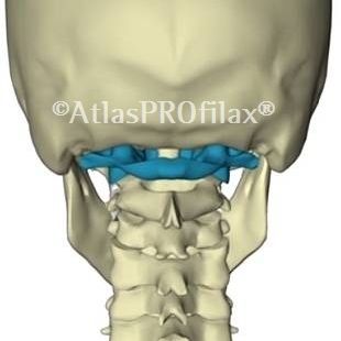
    The upper face or glenoid cavity is articulated with the occipital condyle, thus forming the atlantooccipital joint, which consists of a bicondile diarthrosis formed by condyles of C0 - lateral masses of C1 and fixed by capsules, membranes and atlantooccipital ligaments. Its function is the flexion, circumduction, extension and lateralization of the head, with very little rotation. 

    The C1-C2 joint is created between the inferior veneers of the lateral masses of the Atlas and the superior veneers of the Axis (atlantoaxial) or between the anterior arch of the Atlas and the dens of the Axis (atlantoodontoid). It is the transverse ligament of the Atlas that maintains this joint identity. One third of the internal area of the Atlas is occupied by the dense, another third by the transverse ligament and the last one by the spinal cord.

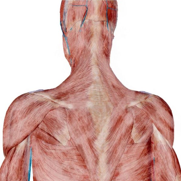
    In the posterior tuber of the Atlas, the posterior minor rectus of the head is inserted; in the anterior tuber and arch, the length of the neck and the anterior minor rectus of the head are inserted while the transverse processes of the Atlas serve as insertion surface for the oblique of the head, lateral rectus, scapula elevator, posterior scalene, cervical splenius, intertransverse of the neck and transverse spinous.

    The Atlas and the human body

    The cerebrospinal system resides inside the skull and the spinal canal. At the cervical level, the spinal canal houses the spinal bulb that emerges through the foramen magnum and makes its transition into the spinal cord. It supports the head and provides protection for blood vessels and nerves that travel through the head. Its wide range of mobility is a consequence of its morphology.

    The cranio-cervical junction has a wide mobile range in flexo-extension and rotation. In the flexo-extension, the occipital condyles move backwards over the glenoid cavity of the Atlas, which also moves backwards. In extension, they move forward, which also moves forward. In rotation, one condyle moves forward while the other moves backward; this is accompanied by lateroflexion.

    The upper segment of the cervical spine, (CO-C1-C2), must vertically balance the cephalic sphere while the lower segment of the cephalic sphere, (C3-C7), must orient the gaze according to the head movements (STP). The negative impact of the lower areas on the head is reduced thanks to the static muscle chain which, by means of the proprioceptors, helps to regulate the dynamics and balance.

    The posterior muscle chain, which begins in the skull and ends at the orthoses, is made up of polyarticular muscles with static function. The suboccipital musculature expresses the articular relationship between the occipital and the Atlas. If there is any irregularity in that joint, the suboccipital muscles initiate an altered transmission of energy to that muscular chain.

    The contraction, irritation and inflammation of the suboccipital muscles leads to the retraction and hypertonia of the posterior chain. These abnormalities vary according to age, the type of fatigue the muscles undergo, and the level of joint stiffness. It can usually result in hyperlordosis, recurvation, and increased supination of the foot, among others.

    The variation in length and viscoelasticity suffered by the aponeuroses makes the tissues prone to high concentrations of waste. Therefore, when there is a mismatch between the Atlas and the occipital, the tension is deposited in the middle septum (connective tissue where the atlantooccipital and atlantoaxial membranes meet) and is transferred to the dorsal raphe (tendon intersection of the upper trapezium, splenius capitis and rhomboid minor). 

    The short muscles of the nape of the neck, which are already susceptible to shortening because they are tonic, will tend to accumulate stress in their fibers and transfer it to the muscular layers that cover them (splenios and trapeziums) as well as the fascias that contain them. This cumulative effect descends through the cervicothoracic-abdominal-pelvic fascia and has a negative impact on the ischiosurales, tilting the pelvis.


    Propaedeutics of the method

    AtlasPROfilax® is born from the combination of biophysics and holistic kinesiology, the latter being a discipline that evaluates the energy, bioelectric and structural imbalances of the body as well as its stress level.

    Holistic Kinesiology does NOT diagnose, it only measures the levels of stress and interference, through techniques such as muscle tone testing. Diagnosis is part of the medical field, kinesiology is not. On the contrary, it corresponds rather to a natural, non-invasive system, which integrates several dimensions of the human being: physical, emotional and energetic.

    Biophysics is a branch of physics that, focusing on biology, applies its knowledge and approaches to living systems, e.g. to describe the behaviour of proteins, enzymes, organic molecules and nerve impulses, etc. Thanks to biophysics, we now have a better understanding of cell interactions, the physical properties of tissues and their responses to certain stimuli, e.g. pressure or vibration.

    Vibration: Propagation of elastic waves that produce deformations and tensions in a continuous environment.

    Pressure: Force that is exerted perpendicular to a surface.

    Kinesiology, auxometry and vibro-pressure

    The AtlasPROfilax® method is based on some kinesiology tests to establish the condition of the atlas vertebra in relation to the occipital bone and the axis. This is possible due to the close anatomical relationship between the tone of the suboccipital muscles and the atlas vertebra. It also uses biophysics to apply a system of controlled mechanical vibro-pressure to the muscles that require it, in order to achieve balance of the muscles, fascia and ligaments in the upper cervical area (atlas-axis). This vibro-pressure is calculated based on auxometric tables (height, weight, age, general health status) and therefore its application is safe.

    Mechanical vibropressure focuses on releasing some of the cervical fascias that are plastically deformed, which is especially relevant because this deformation is responsible for the compensatory effect that the body's longitudinal fascias make. While some discomfort and pain in the head, neck, shoulders and back may be the result of this compensatory stress produced by the deformation of the fascias, their release generates significant reductions in chronic pain (of a non-malignant nature) in more than 85% of cases.

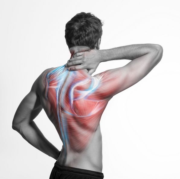
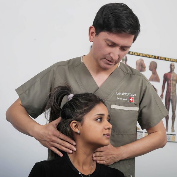
    The final goal of AtlasPROfilax, when carrying out this integration, is to recover the biodynamic balance of the human being as it reduces the stress of the suboccipital muscles. This translates into:

    • Better quality of life.
    • Better quality of sleep.
    • Optimization of posture.
    • Better oxygenation to the brain.
    • Increased concentration.
    • Elevation of energy levels.
    • Increased coordination and balance.
    • Release and disinterference of the brain stem.
    • Improvement of the functions of the neuro-vegetative system.
    • Reduction of the limitations to the articular mobility.
    • Diminution of the intensity of the pain (of not malignant character).

    Theoretical foundations

    In case of a Minor Atlas Intervertebral Derangement, the muscular tests used in the AtlasPROfilax® method usually find hypersensitivity of the cervical musculature, joint asymmetry, movement restriction, trigger points referring to face and neck as well as a good number of muscle shortening at distance.

    The reason why AtlasPROfilax® works specifically on the short muscles of the nape of the neck ( rectus and obliquus of the head) is because these muscles play a decisive role in the body's proprioception (balance - coordination) and nociception (pain signals). This is especially important because, by restoring the balance of these muscles, the myodural bridge is indirectly freed from tension and thus allows the atlas vertebra to regain a proper fit with the occipital condyles.

    This indirect method does not represent any risk because under no circumstances does it directly manipulate the atlas vertebra. The correction of its Cranio-Caudal Deviation and Occipital Condyle Compression is achieved by changing the tone of the muscles, fascias and ligaments involved. The greatest goodness of this is that by applying controlled mechanical vibro-pressure to certain muscles, we obtain a change in tone that is transferred to the atlas through other soft tissues: the ligaments. Furthermore, this correction is preserved over time, unless the person suffers significant trauma to the head or coccyx, e.g.

    Mechanical vibropressure has been calculated based on two laws of biophysics that are useful for fascial work. They are:

    Arndt-Schultz's Law: SWeak stimuli excite physiological activity, moderately strong ones favour it, strong ones delay it and very strong ones block it.

    Hilton's Law: It is related to the capacity of the nerve - which innervates a joint - to innervate also the muscles that mobilize this joint, as well as the skin that covers the joint insertion of these muscles.

    The suppression of the stress of the suboccipital muscles can also result:

    • Less irritation of the posterior cervical ligament. This is directly linked to an improvement in the vascularization of the brain and the preservation of the identity of the pia mater ligaments.
    • Recovery of body balance in people with cervical vertigo.
    • Reduction of the disturbed nociceptive signal of the skeletal muscle, with which painful syndromes such as fibromyalgia can be improved.
    • Desensitization of the C1 nerve root, which allows for improvement of cervicalgia and chronic pain.
    • Decreased levels of fatigue.
    • Drop in hypertone levels in the cervical musculature, which usually improves chronic postural tension.

    Effects of AtlasPROfilax®

    • It results in a natural repositioning of muscles and ligaments, which tends to improve posture and significantly decreases the excess pressure suffered by some particular areas of the body (cervical, lumbar, pelvis and knees). This can benefit especially people suffering from chronic back pain, scoliosis, lumbar disc herniation and hip arthrosis.
    • It frees the Willis polygon from unnecessary compensation by freeing up the correct passage of the vertebral arteries. This restores regularity in the blood flow to the brain and as a consequence, a good percentage of headaches, migraines, dizziness and depressions are usually improved and attenuated. This same fact also affects positively the children's performance at school.
    • The natural diameter of the spinal canal is recovered in relation to the occipital foramen and this leads to a significant reduction in some of the vegetative disorders (such as insomnia and dizziness) and also tends to improve nerve communication from and to the extremities, thanks to which some people manage to recover to some extent the strength they had lost.
    • It releases the dura mater from permanent traction and therefore the irritated spinothalamic and spinocerebellar tracts gradually return to normal. This produces a feeling of greater well-being while significantly improving pain (nociception) and stability (proprioception). This favours those suffering from chronic pain, fibromyalgia and odorous myofascial syndrome.
    • It successfully helps in restoring vital tone and internal balance. However, this kinesiological method does NOT replace medical treatments or medications and does not interfere with them. Far from being an "intruder" of medicine, rehabilitation and physiotherapy, it is a very useful tool because it elevates the quality of life of people suffering from chronic pain and who have previously exhausted all other instances.