Fascia and AtlasPROfilax®

Fascia and AtlasPROfilax®

Technical information for health professionals about the AtlasPROfilax® method

The fascial system

According to Adrej Pilat, one of the great researchers of fascia and creator of myofascial induction, “The body's fascial system forms an unbroken network that controls all the components of our body in different ways. It is not possible to maintain a healthy body without a healthy fascial system. This system should be in functional balance to ensure the body's optimal performance of its tasks. Restrictions on the fascial system and its internal structure create "discomforts" that interfere with the proper functional performance of all body systems. The fascial system may be under excessive stress or it may be too distended; in both situations, body function is affected. This behavior can be compared to three ways of lying down in a hammock: too tight, too loose or perfectly balanced between two trunks; only in the last one is the body comfortable”.

Definition

We define the fascia as a sheet of connective tissue that surrounds the muscles, bones, organs and all other structures of the body, both on a macroscopic and microscopic level. If we speak of a fascial system and not of fascias it is because no fascia is independent of the others, but they are interconnected with each other in such a way that they create a unique network that extends throughout the whole organism and relates that every cell, every tissue and every organ of the body. To show an example, in the muscle, there is a fascia that wraps each muscle cell, called endomysium, each bundle of muscle fibers is wrapped by another membrane called perimisium and the whole muscle by another called epimisium. In reality, all three are extensions of the same fascial tissue that connect them to each other and to the rest of the muscles and other organs of the body.

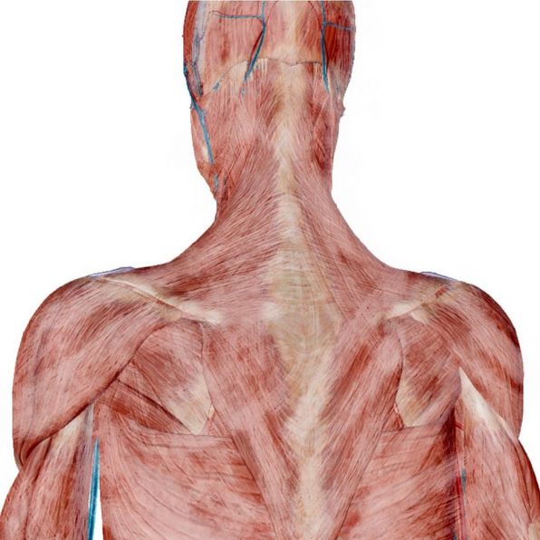
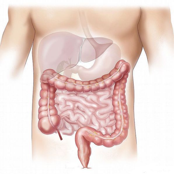
A healthy fascial system allows the different organs to glide over each other to allow a correct functional development. For example, the fascia of a muscle allows it to slide over the other muscles during contraction. The sliding of the pleura allows the expansion of the lungs and the rib cage by allowing the structures to move over each other. When the fascial system is unhealthy, mobility restrictions occur, which, due to the continuous nature of the fascial system, may affect the function of other organs located at a distance.

Functionally, the fascial system also includes the meninges, and the dura mater is the most important membrane due to its characteristics (greater thickness and less elastic capacity). Some authors call the meninges the Internal Fascial System and the rest of the fascial system the External Fascial System.

    Composition of the Fascia

    On a histological level, when we talk about fascia we're talking about connective tissue. This tissue accounts for more than 15% of body weight and contains a quarter of the water in the body.

    Connective tissue is responsible for maintaining the difficult balance between stability and elasticity in the body.

    In terms of its composition, the main component is the fundamental substance. The remaining components (cells and fibres) are found in this gel, which contains a large amount of water. The fundamental substance together with the fibres forms what is called the extracellular matrix.

    The basic substance is basically composed of proteins called glycosaminoglycans (GAGs), which can attract large amounts of water, which is necessary to maintain the mechanical properties of the connective tissue.

    he other component of the extracellular matrix is formed by a series of fibres: elastin, collagen and reticulin.

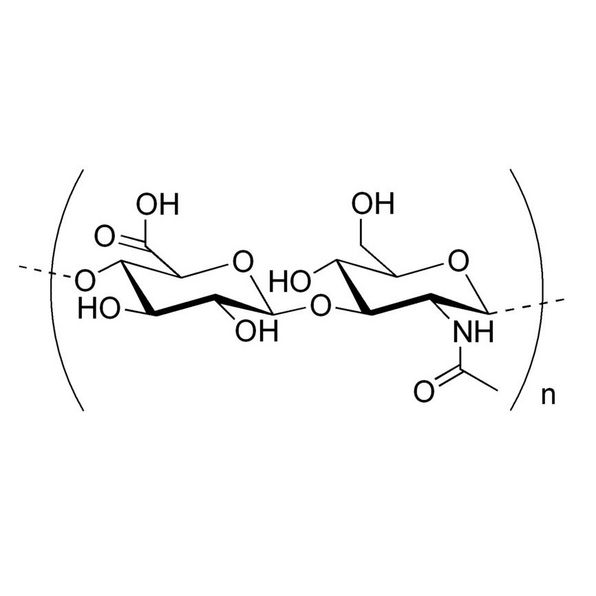
    The third component of the connective tissue is the set of cells, divided into two groups: fixed cells and free cells. Among the former are fibroblasts, whose function is to produce elastin and collagen, and adipose cells, whose function is to store lipids and then release them into the blood for energy.

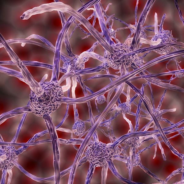
    As for the free cells we can find macrophages, which "eat" the damaged cells and other waste substances when there is an injury and participate in the process of repairing damaged tissue, and mast cells, which participate in the task of renewing the fundamental substance, since their function is to release certain substances necessary for the repair or healing process, particularly produce histamines, serotonin and heparin in the early stages of the inflammatory response.

    Regular dense tissue has a higher proportion of collagen fibres to the detriment of the amount of fundamental substance. In the tendons the distribution of collagen fibres is parallel, while in the ligaments it is more irregular and depends on the mechanical needs of each ligament.

    It is also important to remember that in the fascial tissue there are smooth muscle cells, which give the fascia the ability to contract. According to different authors, the fascia could respond to different traumas of both physical and physical origin by contracting and thus creating a hypomobility.

    In addition to muscle cells, fascia is also rich in nerve endings and sensory receptors such as Golgi, Pacini or Ruffini receptors, which make the fascial system a fundamental part of the proprioceptive system.

    Classification of fascias

    On a general level we find the superficial and deep fascia, with the deep fascia specializing in a number of different structures.

    The superficial fascia is a subcutaneous fascia, in fact it adheres to the skin, which has a different density in each part of the body, being more dense in the extremities and less in the head and trunk.

    The deep fascia integrates the microscopic and macroscopic fascial systems and gives the different organs and systems of the body the quality of being a unit both at a structural and functional level. The deep fascia is denser than the superficial fascia and has a higher proportion of collagen in its extracellular matrix.

    Within the deep fascia, different levels are established:

    • Myofascia: There are different levels of fascia within the myofascia, since in addition to the external fascia of each muscle, the epimysium, each bundle of muscle fibers is wrapped by the perimysium and each muscle fiber or cell by the endomysium. Let us not forget that they are not independent structures but interconnected with each other and with the bone.
    • Viscerofascia: By this term we refer to the different envelopes of the viscera, such as the peritoneum, pleura or pericardium, which are also linked to the myofascia and play a fundamental role in visceral function and in body statics and dynamics.
    • Neurofascia: The structure of the different fascial layers of the nerves is quite similar to that of muscle. In this case, the outermost layer that surrounds the entire nerve is called the epineurium. Each bundle of nerve fibers is covered by the perineurium and each fiber is inside the endoneurium.
    • Meninges: The meninges are considered part of the fascial system (internal fascial system) because of their structure and function, although the one that has the greatest mechanical impact is the dura mater. We distinguish between the cranial dura mater, strongly anchored to the bones of the skull, and the spinal dura mater, which is anchored to the foramen magno, high cervical and sacral segment and coccyx.
    • Myodural or vertebrodural bridge: It consists of a sheet of connective tissue that joins the spinal dura mater with the epineurium of the minor rectus posterior muscle of the head, thus establishing a junction point between the internal fascial system and the external fascial system. This junction bridge can transmit the stresses between the internal and external fascial system. This junction also allows the lesser rectus posterior muscle of the head to take on an important mechanoreceptor function that controls the position of the head and informs the CNS of the stresses experienced in both fascial systems.

    Functions of the fascias

    Just as the fascial system relates to all the structures of the body, its functions also vary greatly, and among them are:

    • Providing mechanical protection to the various organs and structures they surround
    • Acting as a barrier against pathogens (e.g. dura mater)
    • Allowing different body structures to slide so that movement is possible
    • Separating different structures to form partitions
    • Assisting in wound healing
    • Storing lipids, water and minerals
    • Maintaining blood and lymphatic circulation

    Starting from the existence of the fascial system capable of integrating each structure of the body, whether at a microscopic or macroscopic level, different therapeutic currents are born which have the common objective of understanding the organism as a whole and having a new vision of the pathology, which is based on the macroscopic but also cellular restriction and deformation of the fascial system, in order to develop effective liberation techniques capable of restoring the body's capacity for movement and consequently recovering health.

    Although each of these currents has certain theoretical principles about pathogenesis and therapeutic action, all of them nourish the theoretical principles of the AtlasPROfilax® method, which far from entering into theoretical or dogmatic discussions, has been able to take the essence of each of them and integrate them in order to better understand the great therapeutic potential of the method itself.

    The AtlasPROfilax® method has the virtue of influencing the whole craniosacral system in a very direct way through different pathways: the frequencies of vibro-pressure by soft tissue resonance favor a release of fascial extensions that are projected to the dura mater, with a downward effect that will release occipital and sacral at the same time. Secondly, the disappearance of the Reactive Syndrome of the Suboccipital Muscles, mainly the rectus capitis posterior minor, will release tension from the entire external fascial system. Due to its location and the fascial connections of the area, this muscle will probably have a direct action on the cisterna magna and therefore on the CSF fluctuation.

    The fascia can hardly be understood without understanding the concept of "muscular chains". De Méziéres takes the definition of a muscular chain as a "set of at least two polyarticular muscles (whose origins and/or insertions are intertwined) which are organized by overlapping each other and which are connected by the fascial network, where all the elements are in solidarity". For it there is a posterior chain formed by the deep dorsal muscles that acts continuously against gravity and that tends to retract and to fibrosis, since the fibrotic tissue is more energetically efficient.

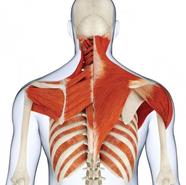
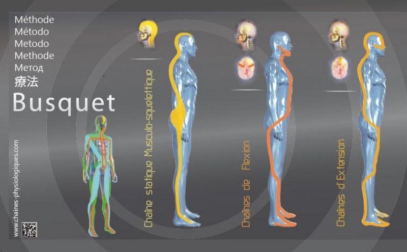
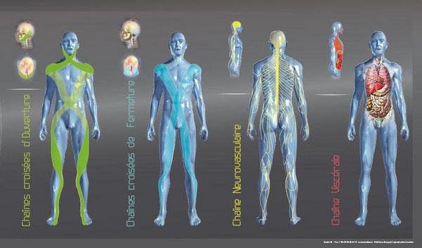
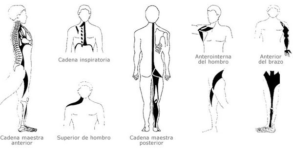
    Leopold Busquets adds some interesting notions such as the inclusion of the dura mater and the visceral fascia. He also establishes the existence of a posterior static chain (PSC), which he considers to be formed by elements of connective tissue such as the cerebral falx, the nuchal septum and the thoracolumbar fascia among others, but he also establishes two other static chains, visceral and neurovascular, thus giving great importance to the connective or fascial tissue as opposed to the muscular tissue. It also considers that there are some dynamic chains involving flexion, extension, opening and closing.

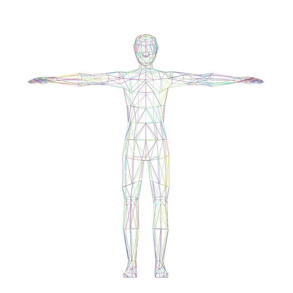
    Images of muscle chains taken from L. Busquets. In yellow the CEP and the neurovascular C. and in red the visceral C., in orange the flexion and extension C., in green the opening C. and in blue the closing C.

    Another of the most renowned authors is Philippe Souchard, creator of the Global Postural Re-education (RPG) which also establishes shortening of the static chains as the cause of the problems of the locomotive system.He underlines the importance of the high cervical segment and its short muscles together with the SCM to maintain the horizontality of the gaze in the face of any imbalance such as scoliosis, etc., when this system is not capable of compensating there is a serious problem.  For disc herniations, arthrosis and other alterations of the locomotive system have their origin in the shortening of the static chains.

    Muscle chains according to P. Souchard

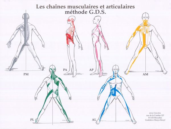
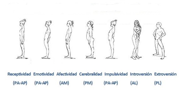
    Other authors such as J. Pilates, Ida Rolf, Thérese Bertherat, Alexander point out various approaches and interrelations of the myofascial system. For example, Godelieve Denys-Struyf, talks about the relationship between mechanics and body statics with psychological aspects. This author states that the function of the body is to express the psychological aspects of the person. She also introduces the term joint chain, as a set of joints that act in a coordinated way in the different movements. The muscle chains sit on the joint chains and are the motor of the joints.

    Muscle chains according to G. Denys-Struyf. In grey the medium posterior chain, in red the posterior-anterior chain, in pink the anteroposterior chain, in yellow the medium anterior chain, in green the posterolateral chain and in blue the anterolateral chain.

    Postural typologies according to G. Denys-Struyf

    Given the above, we can draw different conclusions. Firstly, the existence of static and dynamic myofascial chains, with the static ones being responsible for the posture. These myofascial chains possess a large amount of connective tissue that is more effective than muscle tissue for this purpose.

    Although there are great methodological differences between different authors not mentioned here for practical reasons, there is consensus between all of them on some points. On the one hand, they all agree on the importance of the fascias in the whole system and that shortening one part of the chain has an impact on the whole chain. Furthermore, if we look at the static chains in each of them, they are born or have a critical point in the craniospinal junction.

    From the point of view of the AtlasPROfilax® method we can state that the knowledge of the myofascial chains allows us to understand in a large percentage how and why such important and rapid changes in the posture of our patients occur and why joint mobility is restored and pain reduced within a few days or weeks of the AtlasPROfilax® treatment, even after suffering significant restrictions and deformations. The elimination of the Reactive Syndrome of the Suboccipital Muscles, the decompression of fascial structures with biochemical changes in the atlanto-occipital hinge area releases the muscle chains producing a great physical and psychic benefit on the organism of our patients.

    Fascia and research

    Over the past decade, fascia has been the subject of serious research and has generated numerous scientific books and articles that open up its therapeutic potential. Researchers such as neuroanesthesiologist Werner Klinger, neurophysiologist Heike Jäger, neurologists Carla Stecco and Helene Longevin, from the German University of Ulm, the Italian University of Padua or the Canadian University of Vermont, are leaders in research on the physiology and physiopathology of fascia that may promote new therapeutics in the field of medicine with concrete applications in various pathologies and, in particular, in chronic pain of non-malignant origin. Dr. in Biology Robert Schleip, has been one of the most outstanding and recognized scientists in this field. He leaves us his testimony about the Atlasprofilax method after having received its application. To see the video click here.

    We recommend the viewing of this documentary about fascia made by the German channel Deutsche Welle. To see the video click here.

    Bibliography

    • Andrzej pilat , 2003. Terapias miofasciales: inducción miofascial.  624 págs. Mcgraw-Hill/Interamericana de España.  Isbn: 9788448605599
    • W. Myers, Thomas (2011). Vías anatómicas: meridianos miofasciales para terapeutas manuales y del movimiento.
    • Fernández Pérez, Antonio Manuel. Efectos neurofisiológicos, psicoinmunológicos y psicológicos a corto plazo en sujetos sometidos a técnicas de inducción miofascial.
    • Paoletti, Serge (2004). Las fascias. El papel de los tejidos en la mecánica humana.
    • Agamez Triana J, Arenas Quintana B, Restrepo Barrero H, Rodríguez Giraldo JE, Vanegas García JH, Vidarte Claros JA. Cuerpo movimiento perspectiva funcional y fenomenologica. Universidad Autónoma de Manizales; 2002.
    • Moore KL, Dalley AF, Agur AMR. Anatomía con orientación clínica. Buenos Aires [etc.]: Médica Panamericana; 2007.
    • Paoletti S, Sommerfeld P. Las fascias: el papel de los tejidos en la mecánica humana. Barcelona: Editorial Paidotribo; 2004.
    • Smith Agreda V, Ferres Torres E. Fascias: principios de anatomo-fisiopatología. Barcelona: Paidotribo; 2004.
    • Mancuso P. El Sistema Fascial. 2008.
    • Day JA, Copetti L, Rucli G. From clinical experience to a model for the human fascial system. J. Bodyw. Mov. Ther. julio de 2012;16(3):372-80. 8. Findley T, Chaudhry H, Stecco A, Roman M. Fascia research--a narrative review. J. Bodyw. Mov. Ther. enero de 2012;16(1):67-75.
    • Kumka M, Bonar J. Fascia: a morphological description and classification system based on a literature review. J. Can. Chiropr. Assoc. septiembre de 2012;56(3):179-91.
    • Stecco L, Schleip R. Manipulación fascial: parte práctica. Caracas: Amolca; 2011.
    • Stecco, C. Functional Atlas of the Human Fascial System. ISBN13: 978-0702044304
    • Findley TW. Second international fascia research congress. Int. J. Ther. Massage Bodyw. 2009;2(2):1-6.
    • Findley TW. Fascia Research from a Clinician/Scientist’s Perspective. Int. J. Ther. Massage Bodyw. 2011;4(4):1-6.
    • Findley T. Fascia research 2012: third international fascia research congress. Int. J. Ther. Massage Bodyw. 2010;3(4):1-4. 38
    • Thomas W. Myers (Foreword), Ph.D. Schleip, Robert (Foreword), Ph.D. Findley, Thomas W., M.D. (Afterword).  Fascia - What is is and why it matters
    • Merino R, Mayorga D, Fernandez E, García R. THE EFFECT OF SOLE SELF-MASSAGE ON THE EXTENSIBILITY OF THE BACK MUSCLE CHAIN IN RIATHLETES. A PILOT STUDY. J. Sport Heal. Res. julio de 2010;3:17-26.
    • Willard FH, Vleeming A, Schuenke MD, Danneels L, Schleip R. The thoracolumbar fascia: anatomy, function and clinical considerations. J. Anat. diciembre de 2012;221(6):507-36.
    • Jiménez Díaz JF. Muscular injuries in sport. Int. J. Sport Sci. abril de 2006;2(3):55-67.
    • Cardero Durán M ángeles. Lesiones musculares en el mundo del deporte. Rev. Ciencias Deporte. enero de 2007;4(1):13-9.