Switch language or website
Switch Language
Other countries and websites
AtlasPROfilax® is available around the world in many languages. Please select your region to switch to another website.
AtlasPROfilax® is available around the world in many languages. Please select your region to switch to another website.
One of the most immediate and interesting changes after the application of the AtlasPROfilax® Method is the recovery or improvement of an orthogonal cephalopodal axis, normalizing the curves of the spine in case of hyperextensions or rectifications of the spine. This leads to an improvement in cases of head anteriorization, shoulder anteversion, hyperciphosis and hyperlordosis (cervical and/or lumbar) and . The reflection of this postural change, which remains unchanged over time in a very high percentage of patients, can be read in stabilometric measurements.
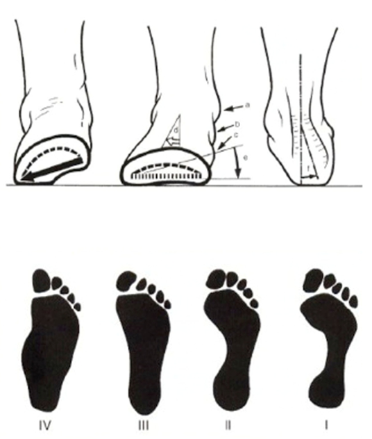
The position of the foot is a reflection of the postural health of the human being. An important distortion and asymmetry in the support of both feet is a factor to be taken into account when looking for injuries or mechanical dysfunctions at the level of the ankles, knees, spine and, therefore, the suboccipital area governed by the head joints and its myotomes.
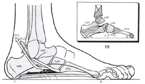
The foot is a relatively rigid structure. It consists of 33 joints and 26 bones grouped together to form a dome that extends from the calcaneus to the distal ends of the 5 metatarsals. The form and plantar curvature is modified so that the foot adapts to the land and, thus, to create a system of cushioning between the ground and the leg that is elastic and easy.
Dr. Pablo Damián García graduated from the Universidad de la Plata (Argentina) as a medical doctor. In addition to his specialization, Dr. Garcia ventured into alternative medicine and has a center where he performs AtlasPROfilax® and other alternative therapies in the city of Bahia Blanca, Argentina. See Dr. Garcia's full profile by clicking here.
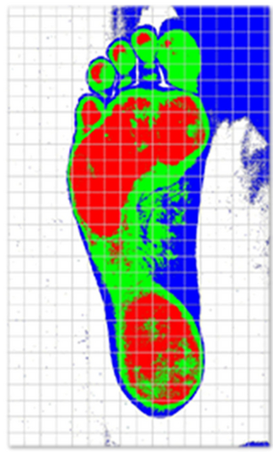
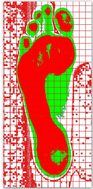
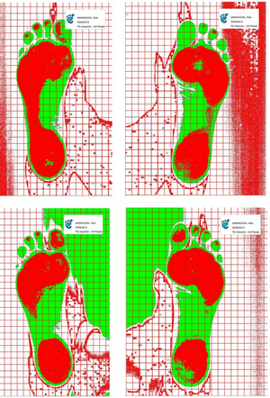
The medium static pressure (MSP) is an algorithm that calculates the average pressure value from the colored surface selected with the footprint index, the total area of the foot and the patient's weight data entered in the personal file. The Footprint Index (FPI) is a number that indicates what percentage of the total foot area is supported with the same pressure. The program uses the following criterion: it marks in red only those areas of the image that are above the selected pressure level. This pressure level is defined by the operator. Finally the program calculates the percentage of the coloured surface in relation to the total surface of the foot.
Since 2012 a study is being developed in more than 350 patients under the direction of Dr. Pablo Damian Garcia, physician and specialist in AtlasPROfilax®. The study is based on observing the change in the scanned footprint of patients undergoing AtlasPROfilax® treatment before and after application. The day of the treatment the patient's foot is scanned, the treatment is performed and the patient is scanned again with the same parameters two months later during a control after the application of the AtlasPROfilax® Method.
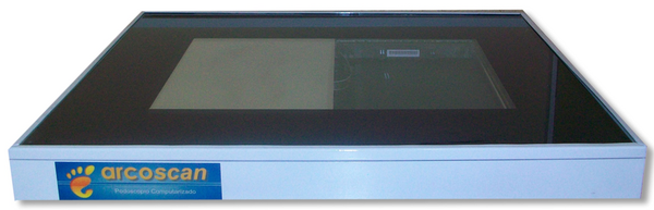
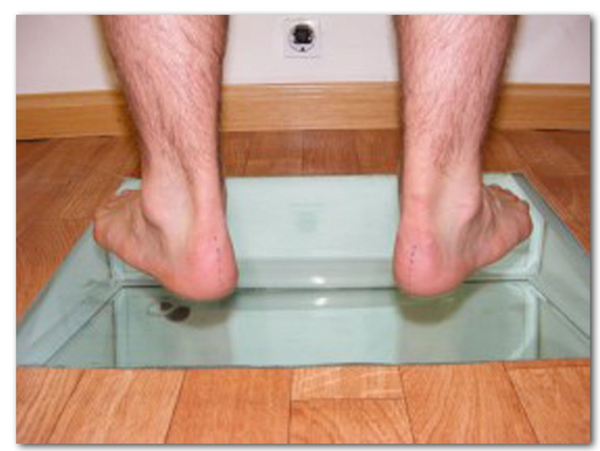
The performance of the podography is simple. The patient, barefoot, is placed on top of the scanner with his arms hanging down at his sides and his feet facing forward.
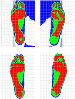
Patient 1: Jorge S. (Male, 43 years old)
The symptoms before therapy were constant cervical and trapezium pain. Pain in elbow and right wrist. Morning low back pain. Pain in big toes. Tension headache twice a week. Morning fatigue that continues during the day.
Upper image: podography BEFORE therapy.
Lower image: podography 2 months AFTER therapy.
Note the improvement in the support of the left foot (to the right of the image). Before therapy the red surface is not well covered. After AtlasPROfilax® the symmetry of the support of both feet is much more balanced.
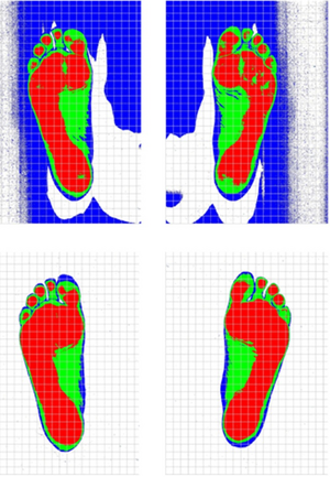
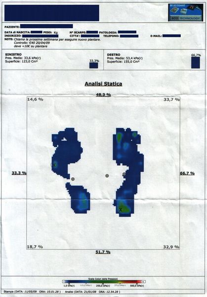
Patient 2: Adriana P (Female, 43 years old)
Adriana suffered from pain in her neck, back, right omalgia and bilateral epicondylitis. As a curiosity, we will add that Adriana suffered from chronic foot pain that disappeared just after the AtlasPROfilax® therapy was performed.
Picture above: podography BEFORE therapy.
Lower image: podography 2 months AFTER therapy.
You can see as in the image below, two months after therapy, the image of both feet is practically mirror-like, unlike what happened before therapy.
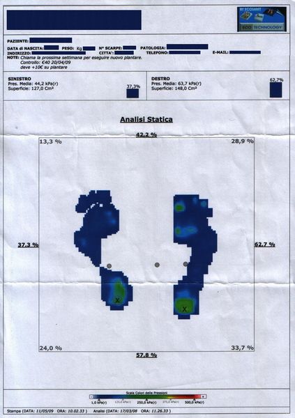
Patient 3: Ana M. (Female, 39 years old).
Ana suffered severe cervicalgia with multiple contractures and pain in her back and shoulders. Ana suffered from insomnia and pain in her feet. She had her toes overlapping and therefore referred that she could not wear boots.
Upper image: podography BEFORE therapy.
Lower image: podography 2 months AFTER therapy.
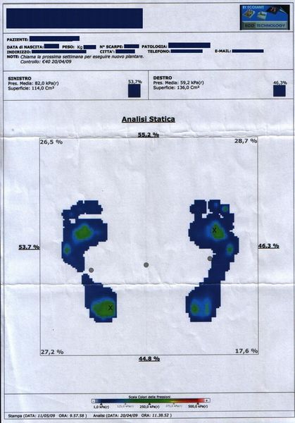
We can see in the lower image a clear improvement in the weight distribution and the balance of the pressure zone in red color much better distributed between both feet than before the AtlasPROfilax® was performed.
We provide here a case of a 28 year old patient with chronic problems of cervicalgia, dorsalgia and lumbago. It refers to kyphotic posture and pains in hip, knee and foot joints.
A significant data of the Atlas correction with AtlasPROfilax® in relation to the change of posture is that the degree of postural relapses is minimal (less than 8%) which guarantees at the same time a much more natural static and biomechanical that improves the quality of life and reduces the risk of muscle injuries or joint wear due to asymmetric overloads.