Switch language or website
Switch Language
Other countries and websites
AtlasPROfilax® is available around the world in many languages. Please select your region to switch to another website.
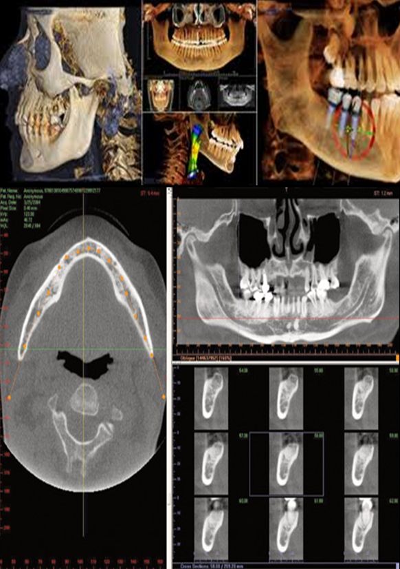
The objective was to check the mechanical changes of the Atlas position in a patient before and after the application of the AtlasPROfilax® Method for Atlas correction.
The patient was treated with AtlasPROfilax® therapy, which consists of highly specialized vibropressure on the short muscles of the nape. Prior to the therapy, the patient had undergone a tomographic scan. Two months later, the patient underwent another tomographic study under the same conditions and according to the same parameters as the first one to determine variations and changes in the position of the Atlas by comparing both imaging tests.
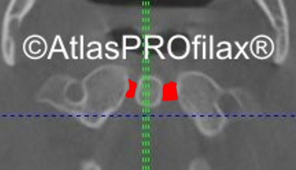
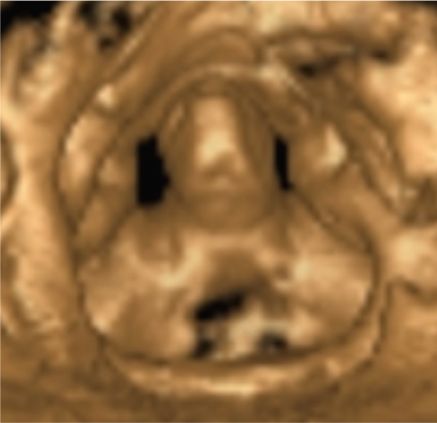
Note in the axial section the rotation of the Atlas with respect to the natural orthogonality. In the coronal section, a clear asymmetry can be seen as only one of the two transverse apophyses of the Atlas is shown (the left one), the other transverse apophyses being relegated by the rotation to a plane that the image does not show. The mechanical position of the atlas is not correct, it suffers from a malrotation with craniocaudal deviation.

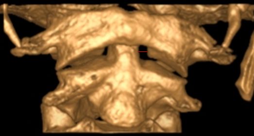
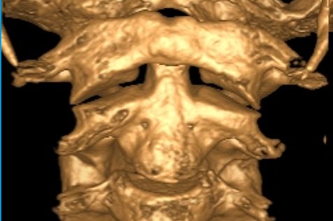
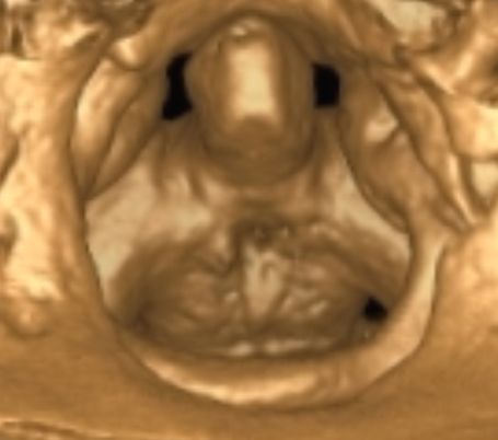
After the AtlasPROfilax® therapy a very clear change in both planes can be observed. The previous malrotation of the Atlas has normalized and disappeared (see blue line). At coronal level the left process has disappeared as the malrotation has normalized. Unlike the previous shot, the coronal section now shows the lateral masses of the Atlas without only one of the transverse processes appearing.

Note how the malrotation of the Atlas, prior to therapy, involves a translation or displacement of it creating an asymmetry between the distance of the lateral masses of the Atlas with respect to the odontoid process. The distance is almost double from one side to the other.
Note the change in distance between the odontoid process and the lateral masses in the Atlas. The distance is, after the correction of the Atlas with AtlasPROfilax®, practically the mass and in any case much better than in the image taken before the application of the therapy.
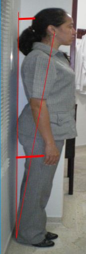
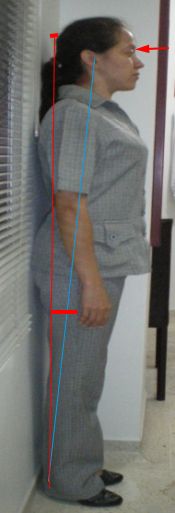
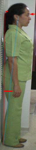
On the clinical side, the patient showed several significant improvements after the correction of the Atlas.
Improved posture:
The Atlas correction changes the center of gravity by improving downward fascial translation and the natural compensatory balance between the various centers of gravity of the spinal segments, the pelvic girdle, and the foot support. See in the example the postural change in the patient.
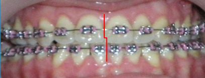
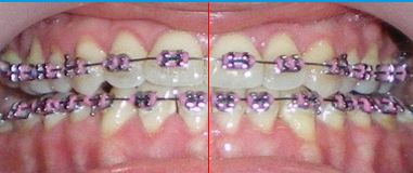
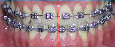
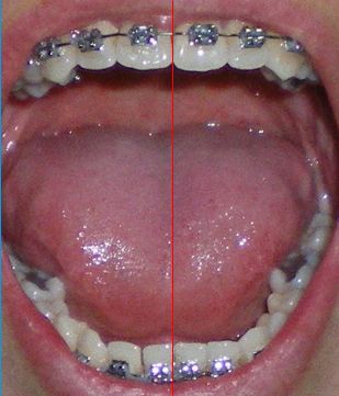
The dental midline and its deviations are an indicator and predictor of temporomandibular joint disorders (see Dr. Gutierrez's study on the "Effect of atlasprofilax® therapy on symptoms related to temporomandibular dysfunction, bruxism and the relationship of dental midlines"). In this case, the patient obtained an improvement in the correlation (before and after therapy) of the dental midline.
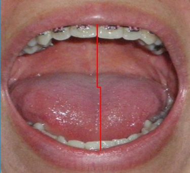
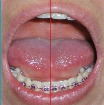
The improvement in mouth opening and midline was also clear after the application of AtlasPROfilax® therapy.